Treatment for Your Child’s Hypoplastic Ventricle: Stage I (Norwood)
Your child has a heart problem that includes a hypoplastic ventricle. The most common treatment is heart surgery. This is often done in 3 stages. But it may need more. Treatment is often complicated. It needs careful management of your child’s health. And treatment does not cure your child’s heart problem. But it can ease symptoms. And it can raise your child’s chances to live a more normal life.
You and your child’s healthcare provider have decided that the benefits of this surgery outweigh any risks. This sheet goes over the surgery that is done during the first stage. Your child’s cardiologist or surgeon can answer your questions. They can tell you more as needed.
The goals of heart surgery for a hypoplastic ventricle
Stage I. Make the 1 working ventricle the main pumping chamber of the heart. This provides stable blood flow to both the lungs and the body.
Stage II. Ease the workload of the 1 ventricle. This reduces the mixing of oxygen-poor and oxygen-rich blood.
Stage III. Separate the circulation of blood in the heart. This means very little oxygen-rich and oxygen-poor blood mix.
Risks and possible complications of heart surgery
These are the risks and possible complications:
-
Abnormal heart rhythm (arrhythmia)
-
Problems in the lungs
-
Problems with the vocal cords and diaphragm
-
Infection
-
Bleeding
-
Problems with the nervous system, such as seizure or stroke
-
Abnormal buildup of fluid around the heart or lungs
-
Death
Stage I: The Norwood procedure
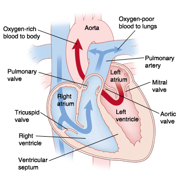 |
| In a normal heart, oxygen-poor blood is pumped to the lungs from the right ventricle. Oxygen-rich blood is pumped to the body from the left ventricle. |
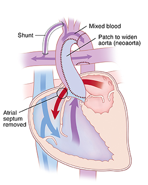 |
| The Norwood procedure uses the right ventricle as the main pumping chamber of the heart. |
The first stage of surgery is called the Norwood procedure. It is often done within the first week after birth. Your child may need to stay in the hospital for 3 to 4 weeks. Your child will need this surgery if the aorta or other left-sided heart structures are too small or missing. This includes hypoplastic left heart syndrome-. The surgery turns the right ventricle into the main pumping chamber. Blood can then be pumped from the right ventricle to the rest of the body to deliver oxygen. The surgery has these steps:
-
Atrial septectomy. The surgeon takes out the wall dividing the 2 upper heart chambers (atrial septum). This lets oxygen-rich blood from the left atrium to mix with oxygen-poor blood in the right atrium.
-
Reconstruction of aorta. The surgeon divides the main pulmonary artery. This artery, along with patch material, is used to make the hypoplastic aorta bigger. The reconstructed aorta is called the neoaorta (new aorta). Blood from the right ventricle can then be pumped through the pulmonary valve to the new aorta. This sends blood from the right ventricle straight to the body instead of to the lungs.
-
Placement of shunt (tube). The surgeon makes a new path to send blood to the lungs. This is because the main pulmonary artery has been used to rebuild the aorta. So, the surgeon places a shunt. It links an artery coming from the aorta to the pulmonary artery. This lets a controlled amount of blood reach the lungs. Sometimes the surgeon may use a method called the Sano method instead of the shunt. In this case, the surgeon places a tube from the right ventricle to send blood straight to the pulmonary arteries. These are connected to the lungs.
When to call the healthcare provider
After any of these surgeries, call your child's healthcare provider right away if your child doesn't seem to be getting better.
Call your child's healthcare provider right away if any of these occur:
-
More redness, draining, swelling, or bleeding at the incision site
-
Fever of 100.4°F (38°C) or higher, or as advised by the provider
-
Trouble feeding, poor appetite, or no weight gain
-
Grouchiness
-
Tiredness
-
Cough that won’t go away
-
Nausea or vomiting that doesn't go away
Call 911
Call 911 if your child has any of these:
Online Medical Reviewer:
Amy Finke RN BSN
Online Medical Reviewer:
Scott Aydin MD
Online Medical Reviewer:
Stacey Wojcik MBA BSN RN
Date Last Reviewed:
3/1/2022
© 2000-2024 The StayWell Company, LLC. All rights reserved. This information is not intended as a substitute for professional medical care. Always follow your healthcare professional's instructions.